Small World Discoveries
by Tony Enticknap - tickspics
Focusing on insects, arachnids, fungus and other small nature subjects from East Dorset and the New Forest ...
Fungal connections (3.3)
The previous two pages in this series regarding 'fungal connections' dealt with specific subjects, starting with fungal mycelium and mycorrhizal fungi, and then saprotrophic fungi, whereas this final page is more of a mixed bag as it covers various topics from asexual fungi and pin moulds, to fungal guttation, lichens and rust fungi.
3.1: Fungal mycelium
3.1a: Mycorrhizal fungi - specifically the ectomycorrhizal 'mushrooms' and their symbiotic relationship with trees
> Mycorrhizal fungi - diversity in a genus (Amanita)
3.2: Saprotrophic fungi
> Brackets and Polypores > Cauliflower fungus > Corticoid (crust) fungi > Pleurotoid agarics
> Saprotrophic agarics > Bonnets > Clavaroid (coral) fungi > Jelly fungi > Cup fungi > Flask fungi
3.3: The confusing world of asexual fungi
3.3a: Mucorales - pin mould
3.3b: Myxomyceticolous fungi
3.3c: Dendrostibella smaragdina
3.3d: Cystofilobasidium macerans - Sap Yeast
3.3e: Fungal guttation
3.3f: Lichenised fungi
3.3g: Pucciniomycetes - rust fungi
Associated information:
Appendix 3A: Ectomycorrhizal fungi partnerships
The confusing world of asexual fungi .....
3.3 (v.2)
Whilst asexual (anamorphic) reproduction occurs across many fungal groups, the most interesting examples that can be seen and photographed in nature are primarily found in the zygomycetes [Mucorales] and some of the synnematoid and cushion-shaped ascomycetes as noted below.
These species, which sporulate through mitotic division by producing identical cloned nuclei from a single parent cell facilitating rapid propagation and colonisation, can be classified in various ways, but in terms of the representative forms featured here are broadly grouped and described as:
Zygomycetes [Mucoromycota] - specifically Mucorales commonly known as pin moulds, which have thin stalked aerial hyphae called sporangiophores that support sac-like spore-holding sporangia.
Hyphomycetes - a class within the now obsolete Deuteromycetes (see below) but still in common use as an informal (uncapitalised) term for asexual fungi which produce conidia on exposed hyphae (conidiophores) either loosely aggregated with a mould-like appearance [formerly classified as Moniliales]; densely adpressed to form a firm stipe with an apical club-shaped capitulum (synnema) that encapsulates the conidia [Stilbellales]; or compacted into cushion-shaped or bulbil-forming structures known as sporodochia [Tuberculariales].
Coelomycetes - another obsolete taxon now treated as a 'form class' for mitosporic fungi that produce conidia in closed erumpent (acervuli) or spherical (pycnidia) fruiting bodies where the spores escape through an opening.
Fungi that are only known from their asexual state were traditionally regarded as the 'imperfect stage' or referenced as
fungi imperfecti and placed in the artificial phylum Deuteromycota [= Deuteromycetes]. Similarly, the sexual spore stage was termed the 'perfect stage'.
The different states are now known as the anamorph being the asexual, often mould-like, reproductive stage; teleomorph (end form) for the sexual, typically fruiting body stage; and holomorph when referring to the whole fungus including both sexual states.
Ascomycetes that have an asexual (anamorphic) state and a known sexual (teleomorph) have traditionally been given separate taxonomic binominals. An arrangement that is confusing enough, but making the connection between the different stages of the same life cycle has further complications especially in the Ascomycetes where, in some cases, the teleomorph is not known or only rarely occurs - a situation which has led to the same species being described in both states without actually realising they're the same species.
Fortunately, advances in mycological techniques over recent years has produced genomic data which has led to many teleomorph binominals being correctly linked to the corresponding anamorph binominal. The only downside is that whilst scientists have been working with these species for some time and are familiar with the names and associated connections, in wider circles having different binominals for the same species has led to misidentification and a growing need for establishing a less confusing system.
As a result, the 'International Code of Botanical Nomenclature' who had previously allowed the use of separate names, decided in 2011 that the arrangement would be abolished and that the teleomorph binominal should be adopted as the official name even if the anamorph had been in longer use. However, scientists pretty much rejected the ruling as the asexual stage of many species had been so widely studied in certain areas, such as agriculture and medical research, that the suggestion of changing established anamorphic names in those particular fields of science would cause even more confusion.
To bring order to the situation, a modified arrangement was agreed in January 2013, known as "one fungus = one name", initiated by the Amsterdam Declaration and implemented in the 'International Code of Nomenclature for algae, fungi and plants' (ICN). The new ruling upheld the previous opinion that the different life stages of the same fungus should not be given separate names, but abandoned the practice that gave precedence to the teleomorph binominal.
As such, mycologist should now be using the earliest published valid name regardless of whether it's the asexual or sexual state, and in situations where two, or sometimes more, binominals were in general use, being able to carry those names forward as synonyms.
It's inevitable that any associated renaming is going to take time to be filter through, so there are obviously going to be situations where outdated or even incorrect names are going to be used, especially as many of the scientific papers relating to these species predate the ruling.
Mucorales - pin mould
3.3a (v.4)
I would never have thought that my recent fascination with some of the more obscure and less popular forms of fungi would extend to mould, but I'm always drawn to the smaller and more unusual species that tend to get ignored, so it's perhaps no surprise that I want to learn more about pin moulds. There aren't that many species that I'm going to be able to find in my local forests and pasture woodlands, but it's definitely a group I want to explore.
In that respect, I should note that this article, like many others that I've written in this series about fungal connections, is primarily for personal reference as I try to obtain a basic understanding of the subject. In this case I simply want to learn enough about pin moulds to be able to distinguish, or at least make an attempt to identify, the different species I might encounter.
Whilst there's a fair amount of information available on the internet regarding zygomycetes in general it's usually associated with biotechnological uses and laboratory applications of agricultural or household moulds, rather than the species I'm hoping to find in nature. In fact it took a surprising amount of time going through scientific papers and various articles to find some of the information I was after and, even now, I still have a few unanswered questions in respect of certain species.
Unfortunately, one of the consequences of trawling through so much data is that certain details and descriptions can become confusing or misleading and, in some cases, contradictory. Everything written here has been checked and cross-referenced as carefully as I can, but it's far from complete as I need more knowledge and certainly more photos as there are a number of species that I've yet to find. As with all of these articles, it can be amended and adapted over time.
The higher-level classification arrangement of the zygomycetes has recently been redefined through phylogenetic analysis, Spatafora et al. (2016), which has left some taxa in uncertain (incertae sedis) position. The upshot of this analysis is that the zygomycetes now comprise two major clades, Mucoromycota and Zoopagomycota, currently defined as phyla, thereby abandoning the traditional Zygomycota. Under this arrangement the Mucoromycota now includes three subphyla - the arbuscular mycorrhizae-forming Glomeromycotina (see article 3.1a); the Mortierellomycotina, which are mainly root endophytes; and the Mucoromycotina, consisting of two primary orders Endogonales and Mucorales, plus Umbelopsidales which separates the genus Umbelopsis with inconspicuous columellae.
Although the group includes a few weak parasites, most Mucorales are saprobes that grow on organic matter such as soil, dung or fruit. They are broadly characterised by having abundant, rapidly growing mycelium typically with long hyphae that develops in the substrate and, through asexual reproduction when conditions are right, produce erect sporangiophores that support sac-like sporangia. They also reproduce sexually through the formation of zygospores, but that's outside the scope of this article which focuses on the general appearance and visible macroscopic features of the anamorphic stage.
Mucorales now comprises thirteen families (previously eleven) with around 55 genera and 260 defined species but, in respect of this article, there are only four families and probably no more than five or six genera, that are likely to be discovered and photographed in the forest or open pasture woodland. Whilst some pin moulds can be identified reasonably easily, the tiny size, variable form and limited visible characters make it almost impossible to distinguish individual species from certain genera. The following is my best effort of trying to describe and identify species I've seen or hope to see.
Mucoraceae - the largest family, although now reduced to 20 genera as Rhizopus, Sporodiniella and Syzgites have been reassigned to the Rhizopodaceae.
Mucor - by far the largest genus in the Mucorales with 70+ accepted species, many of which are clinically important and well documented, but in nature there are only three or possibly four that need to be considered. The most likely is the 'type species' Mucor mucedo, which occurs in all sorts of environments; notably contaminating grain and fruits, damaging cheese or even infecting animals, but in natural habitats, it is most commonly found in soil, growing on dead or decaying organic matter, or more frequently on animal dung, where it is very often the first saprotrophic coloniser.
Growth is normally rapid, initially fluffy, but with sporangiophores soon starting to appear. Erect aerial sporangiophores can grow to between 2-5cm tall or, if prostrate or interwoven in mass as noted below, as long as 15cm. They are predominantly unbranched, although infrequent sympodial branching may be evident, and are pale greyish in colour, later darkening and becoming brown with age. The sporangia, being the terminally-formed closed sacs, are either concolourous or pale beige, turning black at maturity. They typically measure in the region of 150-220µm across.
The interesting thing about Mucor mucedo is that it has different growth forms dependent on conditions and, to some extent substrate, with the primary factors being temperature and light, but also moisture. Unlike other members of the genus, it will grow and produce sporangiophores at temperatures as low as 5°C (as below), but the growth will usually be minimal. Up to 15°C, it will produce shorter, often recurved, sporangiophores than it would when the temperature is in the region of 15-25°C. Optimal growth is around 20°C, but if it starts rising to 30°C or more growth will be inhibited. Another important factor is light, with daylight promoting taller or longer, sparse sporangiophores, whereas more typical, sheltered habitats with poor light will generally produce denser, shorter growth.


Typical growth with a dense mass of sporangiophores from 15-30mm tall - found on a small patch of decomposed matter
on top of a stump in a sheltered, dark habitat during early March with the temperature around 15°C


Sparse short sporangiophores, largest no more than 8mm tall - probably new growth,
on wet, frosty dung (likely deer) in open coniferous woodland with the temperature little more than 5°C

Growing on the remains of a dead snail
When found on fresh horse dung in the middle of a forest track on a warm spring day the identity may well become less certain as the growth is a tangled spreading mass of cobweb-like hyphae (as can be seen in the following photos) rather than tufts of erect aerial sporangiophores with exposed sporangia. Looking closely you may be able to see scattered unbranched sporangiophores and tiny mature black sporangia.


Growing on horse dung as noted above - the example on the right together with an unknown species

Possibly Mucor mucedo, but not dissimilar to the following photos, so not sure!
I've spent ages reading articles about Mucor and Rhizopus to understand the differences and, although I'm reasonably sure that the other examples shown here are correct, I remain cautious when I find fuzzy white masses on dung as both types of mould could be present.
The most important macroscopic characters that distinguish these two common moulds are associated with the mycelium, its origin and branching pattern of the sporangiophores. The mycelium consists of clusters of hyphae, which in Mucor has three different forms: subterranean hyphae, highly branched within or penetrating the substratum; prostrate or aerial hyphae, branched and exposed (without rhizoidiferous stolons); and the sporangiophores, which may be branched or unbranched, but in respect of Mucor mucedo are simple and unbranched,
Other members of the genus to be aware of are Mucor racemosus and Murcor circinelloides which colonise various habitats including dung but, in theory, shouldn't be confused with the common form as they have branched sporangiophores, which are irregularly multiple branched in the former and sympodially branched in the latter, and with much smaller sporangia.
Mucor hiemalis is primarily associated with unspoiled foods, but has also been recorded in soil, litter and decaying plant matter in forests although, as far I can tell, is not found on dung. It has short, unbranched sporangiophores with yellowish-green to brown coloured sporangia.
Whilst Mucor mucedo is regarded as the 'common black pin mould' it is rarely recorded in nature. That doesn't imply that the species is not seen and photographed, just that there are very few verified sightings on either the iRecord or NBN Atlas databases.
Rhizopodaceae - a small family with just three genera including Rhizopus and the monotypic Sporodiniella and Syzgites, all of which weree formerly assigned to the Mucoraceae.
Rhizopus - includes at least eight valid species. Some such as Rhizopus microsporus, Rhizopus oligosporus and Rhizopus oryzae (syn. Rhizopus arrhizus) are particularly well known in connection with the production of certain foods and drinks, or as human pathogens that can cause a severe fungal infection called mucormycosis.
Rhizopus stolonifer, commonly known as the 'black bread mould', is the most prevalent member of the genus and, as far as I can ascertain, the only Rhizopus species that is likely to be encountered in nature as it can occur on fallen fruit and on dung.
Although superficially similar to Mucor mucedo, particularly when seen in mass as both species have a cottony or woolly appearance, Rhizopus stolonifer has a different structure growing as filamentous branching hyphae, but with clustered sporangiophores arising from root-like rhizoids connected by stolons (horizontal runners), whereas in Mucor the sporangiophores are individually borne on aerial or prostate hyphae.
It's difficult to properly see what's going on when you're down on the ground trying to take a close look at the structure of mould growing on horse dung so it's a case of taking different shots from different angles and hoping that sufficient detail has been captured to make an assessment. Often, you'll spot features when looking at the photos later on once you've got them on the computer that were not evident at the time, which can be frustrating if there were further or better details that could have been photographed.

The above photos (set here to magnify rather than enlarge) are close up shots of what I'm currently labelling as an 'undefined species' as there are a few features that don't seem right. Unfortunately we can't see how the hyphae are attached, but we can clearly see are intertwined strands and sporangia that appear to have multiple connection points, which doesn't make sense. We can also see that most of the sporangia are somewhat flattened with a deeply concave (indented) underside, which in isolation is a characteristic feature of Rhizopus when the sporangial walls dry and collapse and start breaking away around the columella, leaving what is sometimes referred to as a "collarette". But Rhizopus has pigmented sporangiophores so, in conjunction with the manner in which the sporangia seem to be attached, there are just too many unexplained details here to even hazard a guess. I hope that if I leave the photos here for reference that one day I'll be able to solve the mystery.
Syzygites megalocarpus [formerly Sporodinia megalocarpus] is a necrotrophic mycoparasitic mould that colonises various species of moribund fleshy mushrooms, such as Amanita, Boletus and Russula. The fast-growing, extensively branched, fuzzy mycelium is initially greyish-white, though often somewhat yellowish due to the presence of carotenoid pigments, later turning bluish-grey to golden-brown.
The mould starts growing from inside the mushroom with the delicate hair-like vegetative filaments arising directly from the substrate. The sporangiophores can grow to around 30-40mm tall when aerial, can be longer when laying across the fruitbody or dangling down from the cap like a veil. Growth is usually quite dense especially around the periphery, but unlike some of the filamentous hyphomycetes that smother their victim, Syzygites magalocarpus usually leaves the host mushroom recognisable.
The sporangiophores are several times dichotomously branched and, once mature, the asexual spore clusters will be contained at the tips within tiny globose sporangia that are just 50-150µm in diameter, initially yellow and then darkening with age. Deep within the mycelium, sexually formed zygospores may also be present, but I'm not sure if it would be possible to spot them when photographing the species in natural habitat.
Syzygites megalocarpus is the sole member of its genus.


Phycomycetaceae - includes just two genera Phycomyces and Spinellus
Phycomyces - three species are listed, although only Phycomyces blakesleeanus and Phycomyces nitens are normally considered; the latter being the originally described form, and the former now regarded as the 'type species' of the genus. They can only be reliably separated by close examination of the spores with those of Phycomyces nitens being somewhat larger, but for general identification purposes they could effectively be treated as the same species.
In natural habitats, Phycomyces is most likely to be found colonising moist organic substrates especially herbivore dung from small rodents such as mice through to larger species like deer or horses. They may also be found on other forms of organic matter in the soil; typically in neutral to slightly acid habitats.
Phycomyces is known for being light sensitive, responding to and growing to light, and when the environmental conditions are suitable will produce impressive, erect, cylindrical, aerial hypha bearing large sporangia. In unrestricted situations the sporangiophores will sprout up from the mycelium at a pretty rapid rate of up to 2mm/hr. They grow from the tip which is initially pointed, but soon swells to form a bright yellow sporangium at which time there's a lull of several hours when the stalk does not lengthen. Once the sporangia are fully formed, growth resumes with the extending sporangiophores twisting in a counterclockwise direction when viewed from above until maximum tension is reached at which point rotation is reversed. They may grow as long as 10cm or more, although the lack of support will usually limit their spread.
As the sporangiophore extends the sporangium starts changing colour from yellow to dark brown or black. This part of the process is happening at different rates so, if the timing is right, it's possible to see some newly formed yellow sporangium mixed in with the mature black ones.
The lifecycle of Phycomyces is well-documented based on laboratory research where optimum growing conditions can be controlled, but in natural habitats the situation is very different as the species is extremely sensitive to light, temperature, wind and even adjacent objects that it will actively avoid. The mycelium is occasionally detected in dung during forest surveys and restricted growth may be found now and again, but erect fruiting growth is rarely observed in the wild as it appears and declines so quickly.
I was rather lucky finding the example seen below which was in the third stage of development after the sporangium have been formed, but before growth resumes and they change colour. Just a few hours either way and it would have looked very different. The second photo shows the species growing on organic matter of some form that was attached to a small piece of deadwood that I'd found partially buried in leaf litter. Although they're out of focus, there are a few yellow sporangia mixed in with the mature darker coloured ones. The identity can't be confirmed, but the thickness of the hyphae and the sheer size of the sporangia strongly suggest Phycomyces.

A lovely fresh bunch of sporangiophores around 5-6cm tall

Muddled restricted growth as noted above
Spinellus fusiger - commonly known as 'bonnet mould', this mycoparasite or 'facultative parasite', is most often found growing on the caps of various Mycena spp. or very occasionally on other small agarics such as Hygrophorus and Collybia.
It's a particularly photogenic mould that I really want to find, but at the moment I can only confirm that it's characterised by dense clusters of erect, radiating sporangiophores that emerge from all over the cap forming a pale brownish-yellow pin-cushion structure that gives the fungus a spiny appearance, hence the generic name Spinellus. The sporangiophores can grow as long as 3cm, each terminating in a dark, spherical sporangia that is 120-300µm in diameter. The texture is fragile and cottony, easily disturbed by touch, wind or rain and, therefore, there's an element of luck involved in finding them at their best. There are a couple of other species in the genus, but as far as I'm aware they do not occur in Britain.
[photos required]
Pilobolaceae - includes just two genera Pilobolus and Utharomyces
Pilobolus - commonly known as the 'dung cannon', is a tiny, coprophilous fungi that has a unique ballistic spore discharge mechanism that ejects the sporangium with incredible force. The spores develop in the sporangia in normal fashion, but to propel the sporangia the stalk swells up with mucus forming a ballon-like vesicle just below the black lens-shaped cap. Once released, the pressure enables the sporangia to be propelled away from the dung where the sporangiophores are growing. They can end up a metre or more away, but it's not done randomly as the sporangiophore is able to control both the angle and direction of fire. I'm not totally sure why, but apparently, they're always projected towards the sun where it is hoped that they will come to rest safely in the grass where they won't get trodden on. It's then a case of waiting, as the life cycle of these species is dependent on the herbivore grazing habits as the spores need to be ingested whilst the cow or horse is eating the grass, but then must pass intact through the animal's gut in order to germinate within fresh dung.
Five species have been recorded in Britain all of which have slightly differently shaped sporangia, but I don't believe that they're distinctive enough to confirm the species except possibly in the case of Pilobus umbonatus which, as its name suggests, has an umbo. I've also got comparative photos of that species alongside Pilobus crystallinus and they look very different so I'll need to do some more research once I've managed to find and photograph them.
[photos required]
Undetermined
Inevitably there are going to be species that I photograph which I haven't yet been able to identify or, indeed, may never be able to identify, such as these two.
The first one is proving particularly frustrating as there are some distinctive features, which I hope will lead to confirmation of
a genus at some point. Whether or not the surrounding mycelia is connected, which I actually doubt, the the mould at the bottom of the mushroom stem appears to be two separate clumps with radiating growth, which is particularly evident in the tiny patch at the top of the stem, and what looks like, long, unbranched, ribbon-like hyphae that are tapered towards the tip. The thick strand of hyphae draped over the top of the deformed stem of the mushroom (likely Lactarius sudulcis, which was found tight to the side of a rotting Beech log) could also provide clues if it's connected to the mould. More importantly, close examination of the hyphae mass on the right clearly shows a dozen or more dark coloured sporangia.
It's been suggested that it's probably just a mass of fungal mycelium, but I'm pretty sure it's not as the structure is just wrong to my eyes. I would really like to know, so I'm going to continue the research, which interestingly has led me to one of the related orders Mortierellales, and possibly the species Mortierella armillariicola as it has a not dissimilar appearance from photos that I've seen on iNaturalist. I obviously need to do more research, but for now I'll leave those thoughts hanging ...
The other photo could be Phycomyes, but I'm not sure, so at the moment it's just here for reference purposes.


Myxomyceticolous fungi
3.3b (v.2)
My interest in this particular group of understudied mycoparasitic fungi is closely associated with the fascination I've acquired for pin moulds and other unusual asexual species, but also with the fact that whilst many people photograph myxomycetes there's a tendency to ignore worn or damaged specimens, or those exhibiting signs of fungal attack.
For this reason alone you rarely see photos of these species and, as a consequence, any associated information is hard to find. A notable exception is the rather attractive Polycephalomyces tomentous which is probably a familiar fungus to anyone that has knowledge of slime moulds even if they haven't actually photographed the species themselves. Typically the knowledge and almost certainly the interest would stop there, which I suppose is understandable as it takes time and patience to photograph these tiny species and very few individuals are going to invest their energies in taking images of white mouldy sporangia that, in most cases, would be unidentifiable without microscopic examination.
I'm under no illusions in that respect, but it doesn't make me shy away from wanting to learn.
From a casual observer's perspective, the various forms of obligate myxomyceticolous fungi that you're likely to encounter could be broadly separated into the following morphological groups:
> synnematoid hyphomycetes which, if my understanding is correct, only relates to two species,
> filamentous 'mould-like' hyphomycetes that typically smother the sporangia, and can be further
separated into two distinct groups being those that attack only non-calcareous myxomycetes, and
those that parasitise calcium-rich species,
> teleomorphic (sexual) ascomycetes,
> other non-specific mycoparasitic fungal species, such as Tilachlidium brachiatum, that have occasionally
been recorded on myxomycete sporocarps but, in all probability, would be no more than a tentative
suggestion based on general appearance.
The classification of these species can be very confusing as the current name of some species has recently been changed, and many have multiple synonyms that have been used over the years. There are too many synonyms to list here, so I've only made note of the most relevant together with the original name (≡) known as the basionym. Strictly speaking, the species name should be followed by the author and date, such as Stilbella byssiseda (Pers.) Seifert, Stud. Mycol. 27: 61 (1985) for example, but that's unnecessary and overkill for a simple article like this.
Polycephalomyces tomentosus (Syn.Blistum tomentosum) (≡ Stilbum tomentosum)
ASCOMYCOTA > Sordariomycetes > Hypocreomycetidae > Hypocreales > Ophiocordycipitaceae
Polycephalomyces tomentosus is presumed to be the asexual (anamorphic) state of Byssostilbe stilbigera (see below), but whilst these two forms may represent different stages of a single (holomorphic) life-cycle they have a different structure and appearance. Polycephalomyces tomentosus is the 'common' form and, from what I understand, the only form that has been recorded in Britain as the much rarer teleomorph is only known from tropical and subtropical regions.
This species has scattered or gregarious, erect white stipitate synnemata with individual structures typically ranging from 250-1100µm tall each with a straight or gently curved, relatively stout unbranched stipe of compact parallel hyphae; arising from a pubescent to dense mat-like layer of hyphae (subiculum), and with each stipe supporting a slimy, globose conidial mass from 50-200µm dia. Microscopically, but possibly visible to some degree in photos, Polycephalomyces tomentosus is characterised by verrucous ornamental cells that cover the entire stipe.
In older literature, reference is made to large oval conidia (characteristic of var.ovalisporum) as well as small conidia associated with the then standard form Stilbum tomentosum. They were later treated as distinct species and described in the genus Blistum, but today both forms are combined, Seifert (1985).
Polycephalomyces tomentosus could be confused with the following species Stilbella byssiseda except that it's almost solely associated with the Trichiales, usually Hemitrichia and Trichia (Oligonema) spp., but also reported on Metatrichia and occasionally Arcyria spp.


Examples of Polycephalomyces tomentosus growing on Hemitrichia / Trichia sp.
Stilbella byssiseda (Syn.Blistum orbiculare, Stilbella orbicularis) (≡ Stilbum byssisedum)
ASCOMYCOTA > Sordariomycetes > Hypocreomycetidae > Incertae sedis > Incertae sedis
This taxonomically unplaced asexual mycoparasitic synnematoid fungi, which has no known teleomorph, is superficially similar to Polycephalomyces tomentosus, but with a different microscopic structure including larger conidia, and although those important characteristic features would need to be properly examined, the species appears more robust. I've yet to encounter this species, so I'll reserve judgement, but I have seen the comparison given as Stilbella = robust (and often fuzzier at the base), whereas Polycephalomyes = fragile.
More importantly, Stilbella byssiseda is primarily associated with Cribraria spp. (especially Cribraria argillacea), with only rare occurrence on other genera such as Fuligo and Didymium. Apart from the odd, possibly questionable record, it is not known to occur on any members of the Trichiales.
Having knowledge of the host species will certainly help when narrowing down the most likely fungus in the following groups, but in respect of these two synnematoids it is widely regarded as a good enough indicator in order to confirm whether it is likely to be Stilbella byssiseda rather than the more frequently encountered Polycephalomyces tomentosus.
Although there are probably around thirty conidial, filamentous 'mould-like' hyphomycetes that that have been recorded attacking and colonising myxomycetes, many are only known from a specific collection(s) from elsewhere in the world and once those are filtered out, there are only a handful of species that are likely to be seen in natural situations. From the information I've managed to pull together, I believe that there are three primary species and possibly four others that could be considered.
They can't be individually identified in the field or from photos, but it's useful to have them listed together with an indication of the host groups that they are known to affect which are included here in taxonomic order.
Aphanocladium album (≡ Acremonium album)
ASCOMYCOTA > Sordariomycetes > Hypocreomycetidae > Hypocreales > Nectriaceae
Forms a loose mass of mycelium that will smother the sporangia of various species within the Cribrariales (eg. Cribraria aurantiaca and rufa); Trichiales (eg. Arcyria cinerea, Oligonema affine, Trichia varia); Stemonitales (eg. Comatrichia nigra, Enerthenema papillatum); Physarales (eg. Craterium minutum, Didymium nigripes, Leocarpus fragilis). No known teleomorph.
Colonies slow-growing, white, composed of fine, hyaline, septate hyphae producing simple, erect, irregularly spaced, awl-shaped phialides bearing conidia that are mostly aggregated in slimy heads.
Gliocladium album (≡ Penicillium album)
ASCOMYCOTA > Sordariomycetes > Hypocreomycetidae > Hypocreales > Hypocreaceae
Recorded on certain members of the Cribariales (eg. Cribraria aurantiaca, cancellata and rufa); Trichiales (eg. Arcyria cinerea, Oligonema affine, Trichia varia); Stemonitales (eg. Comatrichia nigra, Stemonitis fusca); Physarales (eg. Craterium minutum, Didymium squamulosum, Physarum album and leucophaeum). No known teleomorph.
Colonies fast-growing, white, suede-like to downy in texture, with erect, often densely penicillate (tuft-like) conidophores with compacted phialides bearing conidia aggregated in slimy heads.
Verticillium rexianum (≡ Verticillium nanum, var.rexianum)
ASCOMYCOTA > Sordariomycetes > Hypocreomycetidae > Glomerellales > Plectosphaerellaceae
Ubiquitous, and probably the most common filamentous 'mould' on myxomycetes. Recorded on Ceratiomyxa fruticulosa and Lycogala epidendrum, and various members of the Cribariales (eg. Cribraria argillacea, aurantiaca and cancellata); Trichiales (eg. Arcyria cinerea and denudata); Stemonitales (eg. Comatrichia nigra, Stemonitis axifera); Physarales (eg. Didymium nigripes, Physarum compressum). Teleomorph: Nectriopsis rexiana (see below).
Colonies fast growing, white to pale ochraceous, conidiophores usually well differentiated and erect, verticillately branched over most of their length, with whorls of awl-shaped divergent phialides bearing conidia in slimy heads.
Notwithstanding their known host connections, Gilocladium album is recognised as being the most tolerant hyphomycetes in respect of calcium-rich genera such as Physarum, whereas Aphanocladium album is the least tolerant despite associations with other members of the Physarales. On the other hand, Verticillium rexianum has been regularly recorded from various species across all of the main myxomycete orders.


Examples of growing on Hemitrichia / Trichia sp.
(species unknown, but the fine hyphal filaments suggest Aphanocladium album as a possibility)

A distinctive looking example growing on what looks to be Comatrichia nigra
(species unknown, but the form of the conidiophores might suggest Gilocladium album)
For completeness, there are four more hyphomycete species that warrant a brief mention:
Sarocladium bacillisporum (syn.Acremonium bacillisporum) (≡ Paecilomyces bacillisporus)
ASCOMYCOTA > Sordariomycetes > Hypocreomycetidae > Hypocreales > Incertae sedis
This infrequently recorded species is listed as being primarily associated with certain members of the Stemonitales (eg. Enerthenema papillatum and Stemonitis fusca), but also with a few species from other groups such as Cribraria cancellata (formerly Dictydium cancellatum) and Tubifera ferruginosa. No known teleomorph.
Sesquicillium microsporum (syn.Gliocladium microsporum) (≡ Verticillium microsporum)
ASCOMYCOTA > Sordariomycetes > Hypocreomycetidae > Hypocreales > Bionectriaceae
Mainly associated with some of the calcium-rich members of the Physarales (notably Didymium melanospernum and some Craterium and Physarum spp. and also recorded on Leocarpus fragilis).
Although Sesquicillium microsporum has not been linked to a specific teleomorph, it has recently been connected to Nectriopsis and taxonomically reassigned to the Bioectriaceae, and named Nectriopsis microspora.
Verticilium insectorum (≡ Oospora insectorum)
ASCOMYCOTA > Sordariomycetes > Hypocreomycetidae > Glomerellales > Plectosphaerellaceae
Recorded on various myxomycetes including species such as Cribraria cancellata, Hemitrichia calyculata, Comatrichia nigra, Enerthenema papillatum and Stemonitis fusca, but although wide-ranging in that respect, it doesn't appear to be particularly common or note-worthy. No known teleomorph.
Zarea fungicola (syn.Verticillium fungicola) (≡ Acrostalagmus fungicola)
ASCOMYCOTA > Sordariomycetes > Hypocreomycetidae > Hypocreales > Cordycipitaceae
A whitish to creamy coloured mould sometimes with yellowish tones, mostly found growing on cultivated Agaricus mushrooms, but has also been recorded from a few myxomycetes including Hemitrichia calyculata, Metatrichia floriformis and apparently, from some literature, on Stemonitis spp. No known teleomorph.
This final group lists species that have been recorded in their teleomorphic state and, although I may not be able to represent any of them at present, a fair amount of research was required to ensure that the details were correct so are included here for reference.
Six species are noted, but in reality, only Nectriopsis candicans and Nectriopsis violacea are valid.
Byssostilbe stilbigera (syn.Berkelella stilbigera, Stilbella ovalispora) (≡ Hypomyces stilbiger)
ASCOMYCOTA > Sordariomycetes > Hypocreomycetidae > Hypocreales > Claviciptaceae
As previously noted, Byssostilbe stilbigera is widely regarded as the probable, although not yet proven, sexual (teleomorphic) state of Polycephalomyces tomentosus. It is comparatively rare and is more likely to be encountered in tropical and subtropical regions of the world rather than the UK, where it is sometimes found together with new white Polycephalomyces growths surrounding the sexual Byssostilbe perithecia creating a composite appearance.
ASCOMYCOTA > Sordariomycetes > Hypocreomycetidae > Hypocreales > Bionectriaceae > Nectriopsis
Nectriopsis candicans (syn.Nectria candicans) (≡ Hypomyces candicans)
The most frequently encountered myxomyceticolous species of Nectriopsis which, in the teleomorphic state, has a white, floccose (woolly-like) subiculum, with small, superficial, white to creamy-coloured perithecia that have microscopic fuzzy hairs around the pores. It has been recorded from a wide range of host species primarily from the Physarales (eg. Fulgio, and some Diderma, Didymium and Physarum spp.), Trichiales (eg. Arcyria denudata, Metatrichia floriformis) and, to a lesser extent, Stemonitales (eg. Stemonitis flavogenita and fusca). It can be variable in mass, sometimes limited, but typically covering the whole surface of the sporangia or aethalia of the host.
The conidial (anamorphic) state Acremonium sp. is very similar to that found in the following species.
Nectriopsis violacea (syn.Nectria violacea) (≡ Sphaeria violacea)
Although this species has white mycelium it has an overall dull violet appearance as it is densely covered in tiny, partially immersed perithecia pimples, around 200µm diameter, which have a violet to purplish colouration and microscopic hairs forming a fringe around the pores. Only known to colonise Fulgio septica, easily overlooked and rarely recorded.
The whitish coloured anamorph Acremonium fungicola (now treated as a synonym), which has conidia in solitary, hyaline slime heads at the tips of the phialides, may be visible in separate masses amongst the sexual perithecia.
Nectriopsis rexiana (syn.Nectriopsis exigua, Nectria myxomyceticola) (≡ Hypomyces exiguus)
Unlike the previous members of this genus, this form is more usually encountered in its conidial anamorphic state Verticillium rexianum (≡ Verticillium nanum, v.rexianum) as previously featured. In the teleomorphic state it is probably indistinguishable from Nectriopsis candicans without microscopic examination. Associated host species are pretty much limited to Arcyria cinerea and nutans (obvelata), Stemonitis fusca and Fuligo septica.
Two further Nectriopsis species are known to develop on myxomycetes Nectriopsis sporangiicola (syn.Nectria sporangiicola) and Nectriopsis hirsuta (syn.Nectria hirsuta) but, as far as I'm aware, both are from cultivated collections and have not yet been recorded in nature.
Dendrostilbella smaragdina
3.3c (v.1)
ASCOMYCOTA > Leotiomycetes > Helotiales > Tympanidaceae
This very rarely recorded, tiny, rather striking asexual (anamorph) synnematoid fungi has scattered black clubs (synnemata) typically just 800-1200µm tall with a bright blue-green gelatinous head filled with conidia that darkens with age, supported on cylindrical or slightly tapering black stems. Commonly known as the 'teal conifer pin' as it usually occurs on conifer bark, although most of the few confirmed British records for the species, of which there are only seven on the BMS database at the time of writing, are on deciduous wood. The examples featured here were found on the underside of a largish piece of very wet, well-rotted deadwood, which couldn't be determined, but was possibly Hazel.


Cystofilobasidium macerans - Sap Yeast
3.3d (v.1)
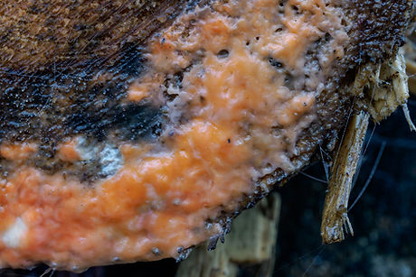
Cystofilobasidium macerans [Tremellomycetes > Cystofilobasidiales > Cystofilobasidiaceae] is the sexual (teleomorph) yeast state of Cryptococcus macerans [Tremellales > Cryptococcaceae], which combines with bacteria and certain types
of fungi to form the slimy, orange, often bubbly substance that is occasionally seen oozing out of tree wounds.
The appearance of this slime, commonly known as Deer Vomit or 'orange-goo' which has no connection with slime mould
per se [Myxomycetes], is typically associated with cooler, wetter conditions during late winter or early spring when sap and nutrients that have been stored in tree roots are transported to the branches to enrich new growth. The sap comes up from the roots under pressure and if the tree has any cuts or wounds that are open to the elements there is likely to be seepage. Similarly if a tree has been felled, as the roots may still be alive and will continue to pump the sap in to what's left of the tree, which is why it is often seen on stumps or on the ends of cut logs.
Sap, or more specifically exuded sap that has been infected with bacteria, attracts wild yeast which interacts with the primary coloniser, which is usually the Ascomycete fungus Fusicolla merismoides, formerly genus Fusarium, [Sordariomycetes > Hypocreales > Nectriaceae]. In this context the fungus provides the sap with body, whilst the yeast ferments within the sap producing carbon dioxide which creates the characteristic orange bubbles. Some literature attributes the colour to either the fungus or the yeast, but in practice it's a result of the interaction between the two organisms which produces carotene - the same orange pigment found in carrots.
Although Deer Vomit is specifically linked with Fusicolla merismoides, every example that has been properly examined has produced a different DNA sequence suggesting that each specimen is a unique complex of different organisms.
Whilst Cystofilobasidium macerans appears to be a constant element, other fungal pathogen species including Fusarium acuminatum, Epicoccum nigrum and Aureobasidium sp., have been detected. In some cases the fungus and associated microbes are existing in some form of symbiotic relationship, whereas others have been identified as parasitising each other.
The biological process here is way beyond my understanding as a photographer of these species, but it is interesting to learn a little bit about some of these unusual fungal forms.


Confusion arises when common names are replicated or incorrectly applied, and there are a couple of these that regularly come up in articles about this species. The first is that Deer Vomit is also used in the US, and possibly elsewhere, as another variant of Dog Vomit (an accepted alternative of Scrambled Egg Slime) in respect of the slime mould Fuligo septica.
The other is Slime Flux, which is an entirely different form of bacterial disease more formerly known as Bacterial Wetwood
that affects certain trees such as Ash, Elm and Oak. Whereas the 'orange-goo' in the featured form above is usually brightly coloured with a feint sweet or yeasty smell, Slime Flux is expressed as a dark-coloured, often foul-smelling, foamy and watery slime that streaks down the bark. Rather than starting from bacteria within the sap, it emanates from the tree's heartwood or sapwood, with the resultant infected liquid and gasses being forced out of cracks in the trunk. Once exposed to the air it will quickly become further infected by various yeasts and other fungi, at which point it is known as Slime Flux.
At this stage it will also become attractive to various insects, such as flies and ants. Deer Vomit may look unsightly, but it's harmless and doesn't last for long, but Slime Flux is a long-term and potentially damaging condition [photo needed].
Fungal guttation
3.3e (v.1)
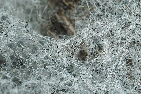
The presence of tiny water droplets clinging to the body hairs of a springtail or transforming a spider's web into a sea of pearls (first photo) would typically add an extra element of interest to almost any macro subject but, apart from the first image, all the 'drops' seen here have been created by the secretion of excess fungal fluids, not from atmospheric moisture causing dew drops.
Informally, these drops of fluid are often described as 'mushroom sweat' but the correct term is fungal guttation. The liquid is a build up of excess absorbed water coupled with various enzymes and compounds that the fungus needs to clear during periods of rapid growth and/or due to external conditions. For most of the time fungi invisibly seep this waste matter through their pores, but too much absorption increases pressure such that it's squeezed out.
These droplets are most likely to be seen on the fertile surface of wood-decay polypores and crust fungus, rather than on agarics or boletes, but also on some other species including a few where the guttation is actually a distinguishing feature of the fungus. And, as well as being found on the fruiting bodies, these droplets can also form on mycelium as the final two photos, or on aerial pin mould hyphae. The exudes are usually clear, milky or yellowish in colour, but darker, orange, red or brown drops are not uncommon, especially during drier periods when the liquid is less diluted than it is in wet conditions.
As a point of interest, all of the droplets in the following photos were found on fungi or mycelium that was hidden away on the underside of deadwood, that was mostly relatively large logs that I'd temporarily rolled over to check what I could find.

Dew drops in a spider's web for comparison


A single large, dark coloured blob of guttation on a polypore

The different colours and reflections that you can capture when photographing guttation can produce stunning images


I don't know the identity of this fungus, but the amber-coloured blobs of guttation certainly add an extra something to the photos


A couple of rather nice examples showing numerous tiny drops of exuded liquid spreading across mycelium
Lichenised fungi
3.3f (v.1)
The vegetative body of lichenised fungi (lichens), called the thallus, is composed of two, or sometimes more, dissimilar organisms that form a mutually beneficial (symbiotic) partnership. The primary element is fungus which typically makes up 80% or so of the body, with the partner most often green alga and/or cyanobacterium. The fungus provides support and can absorb moisture from the air, but as they do not have roots like plants depend on the alga and cyanobacteria which possess the green pigment chlorophyll to produce carbohydrates through photosynthesis.
The fungal component is called the mycobiont. Virtually all lichens have an ascomycetous mycobiont, but there are a few basidiomycetes that are lichenised, notably some agarics of the genus Lichenomphalia (as featured below) and a few small clavarioids in the genus Multiclavula. They are classified utilising the scientific name of the fungus.
The photosynthetic partner or photobiont of most lichens is normally green algae (Chlorophyta) from the genera Trebouxia [class: Trebouxiophyceae] or Trentepohlia [class: Ulvophyseae] and a few others including Chlorella, Diplosphaera, and Myrmecia. However, there are some species, known as 'cyanolichens' where cyanobacterium is the main photosynthetic component, and others that combine with both green algae and cyanobacterium. The most frequent cyanobacteria partners are from the genus Nostoc [class: Cyanophyceae]. The colour of the lichen is largely determined by the photobiont rather than the fungal element even though it's the primary component.
Whilst the fungus cannot survive without the carbohydrates produced by the photobiont, and in many cases the algae cannot grow or reproduce on its own, there are exceptions as some algal symbionts are able to live independently. The best and most visible example in woodland habitats particularly is the alga Trentepohlia, which forms orange-coloured populations on tree trunks (as featured below).
Some species of fungi live on the thallus as an obligate parasite rather than as a component part of the lichen. They are known as lichenicolous fungi and should not be confused with lichenised fungi. One of the relatively common and more visible species is featured below.
Lichens are featured separately in their own section, which provides some basic information regarding growth forms and reproduction, as well as 150+ individual species accounts. The woodland collection includes most of the species I'm likely to find locally, but in terms of this series of articles there also a number of lignicolous and corticolous Cladonia species that will regularly be seen on deadwood and growing on trees. A few common [Lecanoromycetes >] examples are shown below.

Graphis scripta s.lat
Ostropales > Graphidaceae

Physcia tenella
Calicales > Physciaceae

Flavoparmelia caperata
Lecanorales > Parmeliaceae

Xanthoria parietina
Teloschistales > Teloschistaceae

Evernia prunastri
Lecanorales > Parmeliaceae

Ramalina farinaceae
Lecanorales > Ramalinaceae
Lichenomphalia umbellifera (syn. Omphalina ericetorum)
BASIDIOMYCOTA > Agaricomycetes > Agaricales > Hygrophoraceae
The members of this genus live in mutualistic symbiosis with green algae, forming a grainy or leaf-shaped thallus from which the fruitbody arises. Lichenomphalia umbellifera is a deeply decurrent gilled omphalinoid fungus with a tan-coloured umbilicate cap that can become very deep and funnel-shaped. The margin unfurls with age and often becomes pleated or scalloped, making the cap look slightly veined. Typically occurs on moss-covered fallen trunks or logs in damp woodland, and could be easily overlooked as just another small brown mushroom.

Lichenomphalia umbellifera
Trentepohlia spp.
CHLOROPHYTA > Ulvophyceae > Trentepohliales > Trentepohliaceae
Trentepohlia is a genus of filamentous green algae, which is symbiotically associated with lichens as previously noted, but also exists as a free-living organism on the damp, shaded side of tree trunks and other substrates. The 'type species' of the genus and the most widespread form in Britain is Trentepohlia aurea. It consists of macroscopic small bushy tufts which range in colour from golden yellow to orange-brown; the colour resulting from the carotenoid pigments in the algal cells.
In combination with fungal hyphae, they form another defined type of lichen known as a 'protolichen' where they are the phycobiont partner. This form is particularly associated with the crustose 'script lichens' of the genera Arthonia, Graphis, Graphina and Opegrapha.
There are numerous other free-living algal photobionts, but the majority are microscopic species which have been recorded from soil.


Green Algae - Trentepohlia aurea
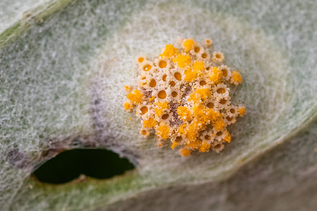
Erythricium aurantiacum (syn. Marchandiobasidium aurantiacum)
BASIDIOMYCOTA > Agaricomycetes > Corticiales > Corticiaceae
Asexual (anamorph) parasitic lichenicolous fungus that forms tiny grainy, pastel orange bulbils up to just 150µm diameter, occasionally forming basidia, but not conidia. Can occur on various lichens, but especially on Physcia and often spreading to the thallus of any adjacent species.

Erythricium aurantiacum (example of a lichenicolous fungus)
Pucciniomycetes - rust fungi
3.3f (v.1)
I was in two minds whether to include an article on rust fungi as I've only photographed a couple of species, which were actually found in grassland rather than woodland habitat, but on the other hand Pucciniomycetes is one of the primary divisions (classes) of Basidiomycota so it should be mentioned if only briefly. It's a very large and diverse group that contains around twenty families of rust fungi in five orders.
The Puccinials is by far the largest order representing, on a worldwide basis, around 7,000 species, the majority of which are in the Puccinia genus. I have no idea how many occur in Britain and apart from taking the odd photo if a particular species has caught my attention, I don't see myself taking more than a cursory interest.
They are obligate plant pathogens that cause damage to various degrees, some severely, but rarely to the point of killing the host. Each species has a range of hosts and, in fact, can infect two different species, during their life cycle, but they cannot be transmitted to non-host species. Beyond that it gets very complicated, so I'll simply close this article off with a couple of examples including Puccinia cnici-oleracei, which has to be one of the most attractive species you could hope to see if found at this stage of development.

Puccinia cnici-oleracei [Pucciniaceae]
on Melancholy Thistle [Cirsium heterophyllum]

Triphragmium ulmariae [Sphaerophragmiaceae]
on Meadowsweet [Filipendula ulmaria]
Puccinia cnici-oleracei [Pucciniaceae]
on Melancholy Thistle [Cirsium heterophyllum]

Triphragmium ulmariae [Sphaerophragmiaceae]
on Meadowsweet [Filipendula ulmaria]

Melamspora rostrupii [Melampsoraceae]
on Dog's Mercury [Mercurialis perennis]