Small World Discoveries
by Tony Enticknap - tickspics
Focusing on insects, arachnids, fungus and other small nature subjects from East Dorset and the New Forest ...
Myxomycetes - slime mould
With ongoing improvements in the design and capabilities of cameras and macro lenses, tiny organisms such as slime moulds that were previously outside the scope of most photographer's equipment, have now become popular subjects. If you have the inclination, patience and skill to capture minute detail across multiple frames that can later then be blended together in special software, you can certainly produce very useable photos of these species. In fact, there are few individuals that have taken slime mould photography to a whole new level, producing beautiful images with incredible detail. Many other macro photographers, including myself, have tried to capture similar detail, but for one reason or another struggle to produce similar results.
I love a challenge and certainly want to spend more time photographing slime mould, but the biggest problem I have is finding them in the first place as it's not just a case of turning over a piece of deadwood. The habitat, conditions and time of year are important, and that's something I need to work on. Hopefully, in time, I will have sufficient material and knowledge to put together a proper album on the main part of this website, but for now I'm going to use this page as a personal learning zone.
I'd originally planned to include a short article for slime mould that was going to be tacked onto the end of 'fungal connections', but with so much to learn, and with so many notes and bits of information that I want to be able to refer to, I'm now going to build on this series of article adding new species and amending detail as needed.
4.1: Classification
4.2: Plasmodium
4.3: Fruiting bodies and terminology
4.4: Ceratiomyxales > Ceratiomyxaceae
4.5: Cribrariales > Cribrariaceae
4.6: Reticulariales > Reticulariaceae
4.7: Trichiales > Arcyriaceae, and Hemitrichiaceae / Trichiaceae
4.8: Stemonitidales > Amaurochaetaceae and Stemonitidaceae
4.9: Physarales > Didymiaceae, Lamprodermataceae and Physaraceae
Classification
4.1 (v.1)
Although slime moulds are now taxonomically placed within the PROTOZOA (=Protista) rather than Kingdom FUNGI, they are often featured alongside each other due to their not dissimilar appearance and the fact that they are frequently found in the same woodland microhabitats. Once you get to know the species there shouldn't be any confusion, but there are bound to be odd situations when something new is discovered when it may not be immediately obvious whether it's a slime mould or a tiny ascomycete fungus.
My interest is primarily with slime moulds that I can find and photograph in natural habitat, not with collecting or studying under a microscope. I appreciate that identification in that respect will be limited and that many species will either be undetermined beyond the genus or even the family, or will be qualified as 'likely' rather than 'certain'. Obviously, I'd like to put a name to all the species I photograph, but as with many of the 'small world' subjects that I've spent time researching I don't have the time or inclination really to take my interest to the next level where it becomes necessary to prepare microscope slides in order to check spore ornamentation and the like. I'll happily leave that to the experts who have been studying these species for many years.
Having spent a fair bit of time checking local distribution records and the various species that other photographers have been able to find, I've put together a list of around 35-40 species that I would like to photograph. I think it's a realistic target,
but in with everything else I want to find, it might take a while. In the meantime though, I shall start with the few species I have photographed and build from there.
But, before looking at the different forms and trying to get to grips with the terminology and the important features and characters of individual species, it's useful to understand how they are classified. I'm not sure how many species have currently been recorded in Britain, but I have compiled a reasonably comprehensive list of around 180 that should cover anything I'm ever likely to find, even though the majority are rarely recorded, let alone photographed.
They are classified within fourteen (...ceae) families and (39) genera as noted below, which are placed in seven orders separated into three subclass divisions or clades:
PROTOZOA - Protozoans (Kingdom) > AMOEBOZOA (Phylum) > Mycetozoa (Infraphylum)
Eumycetozoa (class) [formerly = Myxomycetes > Ceratiomyxomycetes]
> Protostelia (subclass) [formerly = Ceratiomyxomycetidae]
Ceratiomyxales > Ceratiomyxaceae (1)
Myxomycetes (=Myxogastria) (class) - True Slime Moulds
> Lucisporomycetidae (Subclass) - 'bright-spored clade'
Cribrariidia (Superorder)
Cribrariales > Cribrariaceae (1)
Trichiidia (Superorder)
Reticulariales > Reticulariaceae (3)
Liceales > Liceaceae (1)
Trichiales > Arcyriaceae (2), Dianemataceae (3), Hemitrichiaceae (2), Trichiaceae (4)
> Columellomycetidae (Subclass) - 'dark-spored clade'
Echinosteliidia (Superorder)
Echinosteliales > Echinosteliaceae (1)
Stemonitidia (Superorder)
Stemonitidales > Amaurochaetaceae (6), Stemonitidaceae (3)
Physarales > Didymiaceae (3), Lamprodermataceae (3), Physaraceae (6)
Plasmodium
[article requires more photos]
4.2 (v.1)
It's not always visible, but the first sign of a developing slime mould would be the presence of the plasmodium, which differs from the usually dry, thread-like structure of fungal mycelium (see article 3.1) as it will likely have a viscous, slimy consistency. The appearance, texture and colour vary from species to species and may be also be affected by other factors such as the weather, nature of the substrate or indeed what the plasmodia has been feeding on as algae and lichens may produce a greenish tinge. In most cases though, the colour will be hyaline, milky white, or more often yellow, but occasionally other colours can occur.
Although the actual colour is said to be reasonably consistent for a given species, it does not aid identification at this stage.
In fact, the only time that the plasmodium becomes useful in that sense is if you're able to follow development of the species through to maturity, but even then, in most cases, a sample would still need to be collected for microscopic examination as there are very few slime moulds that can be positively identified from photos alone.
It's pity I haven't got better photos, but here are a couple of examples of early plasmodial growth. In the first photo, taken in rather wet weather during the winter, the plasmodium is a small, very fluid patch that hasn't got any structure, but is moving leaving a trail across the substrate. In the second image, taken in the autumn, the plasmodium has started to form into separate growths that I presume will soon start to develop into fruit bodies [further photos needed to show better formed plasmodia].

Plasmodium (emergent)

Plasmodium (developing)
The largest and most conspicuous form that you're likely to find is phaneroplasmodium (Greek phaneros = visible). In fact, it's probably the only type you're going to be able to see as the other two primary forms aphanoplasmodium (aphanes = invisible) and protoplasmodium (proto = first) are thin and transparent or microscopic.
Phaneroplasmodium is also the most variable and typically the most colourful form. It is especially associated with the Physarales, and some Liceales. It can also be related to the Trichiales, although there is a bit of an overlap here with the aphano type, so in that connection it's usually referred to as the trichiaceous type. In most cases it remains concealed in the substrate usually producing fruit bodies on the surface without actually emerging.
When mature, true phaneroplasmodium is characterised by having an anterior fan-shaped structure with channels of streaming protoplasm that enable the organism to creep across the substrate. Unlike individual protoplasmodium that only give rise to a single sporangium, phaneroplasmodium typically forms multiple fruiting bodies often across relatively large areas.
During unfavourable conditions plasmodium can enter a metabolically inactive dormant state but, at some point, the situation will improve and it will migrate to the surface. Freshly emerged plasmodium typically starts out as an amoeba-like mess that gradually take shape as it ingests bacteria and organic matter whilst slowly creeping across the substrate. At some point though when conditions are right, usually during the night or early morning, it will start producing sporocarps. It's impossible to determine when, but the best chance of finding fresh fruiting bodies is probably on a mild morning after a period of rain followed by a day or two of sunshine.
That said, I have to admit that virtually all of the examples shown here were found by chance on or under deadwood when looking for some of the smaller ascomycete fungi or various invertebrate species.
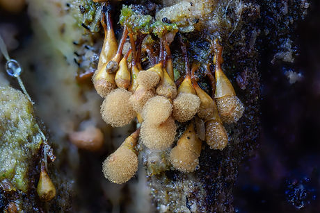
Although the general form and appearance of fresh sporocarps may provide an indication of the species, they should be regarded as unidentified until they're fully developed. Here are two typical examples:
.jpg)
fresh immature sporangia
(despite above comments - possibly Diachea leucopodia)
-2.jpg)
fresh immature sporangia
(despite above comments - likely Hemitrichia / Trichia sp.)
Fruiting bodies and terminology
4.3 (v.1)
Typical slime moulds
Generally referred to as sporocarps, the fruiting bodies of most slime moulds are sessile or elevated structures that are rarely more than a couple of millimetres tall. In the more common stipitate form the stalk supports a spore-bearing sporangium with the enclosed spore-mass protected by an outer covering known as the peridium. In some species the peridium is no more than a delicate membrane that will readily disintegrate or flake away to release the spores, whereas in other species it will have an additional harder and more resilient outer layer that may appear granular, crusty or even powdery. It can disappear completely at maturity or persist in some form after the upper portion has ruptured, which more often than not produces an irregular break, although in a few species it will split neatly around the middle, which is known as circumsessile dehiscence.
The stalk usually extends into the spore-mass terminating in a sterile structure called the columella, which may appear as an extension of the stalk or have a different form. The structure, texture and general appearance of the stalk can vary greatly from one species to another, and may or may not include a cup-like head that supports the sporangium called the calyculus. Around the base of the stalk a thin membrane-like hypothallus may be seen, which is the remains of the plasmodium that keeps the fruitbody attached to the substrate.
The sporangium is even more variable with numerous forms, textures and colours that can appear very different from one stage of development to the next, with many species going through incredible transformations over just a few days or even hours. Microscopic examination of the spore-mass would typically reveal a network of interconnected or free sterile elements known as elaters of various construction and texture, called the capillitium.
Having some basic knowledge of the various parts is useful, although when trying to ascertain the likely identity of a species from field observations and photos alone, you're pretty much restricted to the general appearance and visual features associated with the size and structure of the stalk and the shape, colour and texture of the sporangium. Generally it's the outer wall, being the protective covering of the peridium, that provides the initial, often vibrant, colour in young growth and early development, and then the exposed spore-mass / capillitium once the fruit body is mature.
Alternative forms
Although the majority of slime moulds that I'm likely to encounter will have a sessile or stalked sporocarps as described above, which will typically be in loose or clustered groups where all the sporangia arise from a single plasmodium, a few species develop in other ways.
The first of these alternative forms is a plasmodiocarp, best observed in species such as Hemitrichia serpula, which has a net-like, branched or somewhat ring-shaped structure that, initially at least, retains the general shape of the main veins of the plasmodium. In some situations, short plasmodiocarps can occur, which superficially look like sessile sporangia. I've yet to
see this form [photos needed].
Next is a hemispherical or cushion-like sessile structure called an aethalium, which is effectively a mass of individual sporangia that have fused into a single fruitbody encased in a relatively thick covering referred to as the cortex rather than
the peridium. The most familiar examples of aethalia are in the genera Lygogala and Reticularia (Reticulariaceae), and Fuligo
(Physaraceae), as featured below, but also in Didymium spongiosum [=Mucilago crustacea] (Didymiaceae), and Bredfeldia maxima (Amaurochaetaceae).
The final type is a pseudoaethalium which, as its name suggests, has a composite structure much like an aethalium, but differs in the fact that rather than being completely fused together the individual sporangia remain more or less distinguishable. It is only found in a few species, notably Tubifera ferruginosa [photo needed].
Ceratiomyxales > Ceratiomyxaceae
4.4 (v.1)
Ceratiomyxa fruticulosa is a relatively common and distinctive slime mould that has a different life cycle and structure to any of the other species featured here as its spores are produced externally on tiny thread-like stalks that are attached to the outside of the sporangia. This species can be found at different stages of development and in different forms, including the plasmodial stage when it could appear as a thin white spreading growth, often with a frost-like texture, or as a thicker flowing mass that may end up covering a relatively large area of the substrate. The fruiting bodies typically comprise of numerous erect, simple or branched finger-like columns or as undivided clusters. A well-formed mass resembles coral, hence the common name of 'coral slime'. Generally pure white, but very occasionally pale yellow or pink variations may occur.
Traditionally placed in its own subclass Ceratiomyxomycetidae, but now moved out of Myxomycetes to Protostelia. The genus consists of four species, but only Ceratiomyxa fruticulosa is found in Britain.


Cribrariales > Cribrariaceae
4.5 (v.1)
The Cribraria genus includes small, fragile, attractive species with stalked globose sporangia, lacking a capillitium, and with the spore-mass held together by a net after the peridium disintegrates. They are primarily associated with rotting conifer wood, but may also be found on Birch. Around twenty species have been described, but realistically there are probably only
6 or 7 that are likely to be encountered. I have four on my 'target list'.
Cribraria argillacea - short-stalked, usually tightly packed, but with sporocarps retaining individual identity; peridium soon disappearing leaving a thin dark net, thicker below acting as a cup; spore-mass pale grey; stalk dark brown to black, furrowed; very attractive when mature.
Cribraria aurantiaca - long-stalked, usually in loose groups; peridium soon disappearing, but leaving a well-developed cup and net holding the spore-mass, which is bright-yellow, later fading to ochraceous, although also developing through lovely shades of green and blue; stalk reddish-brown, tapering; plasmodium bright green.
Cribraria cancellata - not quite as attractive as the previous species; long-stalked, in loose groups or in large clusters; peridium disappearing leaving a net consisting of longitudinal thickened bands, no cup; spore-mass reddish-brown, initially paler; occasionally found on Beech as well as conifer wood and Birch.
Cribraria rufa - the type species of the genus, characterised by the netted bright orange to dull red spore-mass when mature, and black stalk; peridium remaining as a cup; also rather attractive during early development, pale bluish-grey to brownish-grey with more distinct net and reducing cup.
Reticulariales > Reticulariaceae
4.6 (v.1)
Lycogala epidendrum has large, 7-9mm dia., scattered to clustered, subglobose aethalia that are initially yellowish-pink, but later acquire a lead-grey to brownish-black surface. There are a couple of other forms, such as Lycogala exiguum, which is much rarer. That particular species is much smaller, initially orange with a scaly cortex, becoming olive brown as they mature. I'm not sure if it's the species featured below, but it is a possibility.
Reticularia lycoperdon is a rather large, non-typical slime mould, that can grow to 8cm or more across. They are initially white to yellowish, but later maturing to a shiny brown-grey colour with a dark brown thread-like 'spore mass'. They can be found growing on deadwood or through mosses as in the example below, or on the bark of various trees, often high up where you might find the similarly shaped fungal species Daldinia concentrica.

Lycogala epidendrum (immature)

Lycogala epidendrum (mature)


Lycogala sp. (see text)
Reticularia lycoperdon
Tubifera ferruginosa - pseudoaethalium (see alternative forms, article 4.3) of pinkish-brown, fading to dull brown, cylindrical sporangia to 5cm x 2cm, and up to 10mm tall; peridium thin, described as having mother-of-pearl reflections; hypothallus white, spongy; plasmodium coloured, orange, apricot or pink; considered common, occurring mostly on rotten conifer longs and stumps, but also on Alder, and more rarely Birch; typically found late summer to early autumn [target species - photos ideally required at different stages of development].
Trichiales > Arcyriaceae
4.7 (v.1)
Arcyria denudata and Arcyria incarnata can appear very similar, which often leads to misidentification. Both species are regarded as common with the former clearly the most frequently recorded of the two nationwide, but with the latter slightly more common locally.
Unfortunately they have a rather fragile sporangium with a spore-mass consisting of a network of fine threads that is easily damaged or dislodged from the calyculus such that, more often than not, you find them in a less than perfect condition.
Arcyria incarnata is typically found in scattered or loose groups rather than packed together. They are much the same size as Arcyria dendudata at around 5mm tall when fully expanded, but with the sporangia having a looser, more delicate capillitium that is only barely attached to the shallow, saucer-like cup. The threads are pale pink to pale crimson with the spores in mass described as rose-coloured, later weathering to reddish-brown if still intact. The stalks are slender and striate, and usually a tad darker than the sporangium, but dark-brown to black around the base.
Arcyria denudata by comparison are gregarious and are generally found grouped together in clusters. The sporangia are cylindrical with rounded or acute apieces and, to my eyes, the capillitium appears course and more densely packed, although when I read the description of the two species the size of the threads is the same. The colour of the spore-mass is variably described as dark pink to bright red. The stalk is dark red to black growing from a thin hypothallus, which is shining reddish-brown with a silvery sheen.
Once the spores are dispersed the empty calyculi usually persist for a while; a good example of this is shown below where there were hundreds of empty caps together with a few remnants of blown sporangia.


Arcyria sp. - possibly Arcyria incarnata
(scattered, sporangia with a loose network of threads and the spore-mass pale pink to rose coloured)


Arcyria denudata
(clustered, sporangia with a tighter network of threads that appear to be more securely attached, and the spore-mass dark pink)

Arcyria denudata
.jpg)
Arcyria sp. - likely Arcyria denudata (immature - clustered)

(tightly grouped in clusters, with bright red spore-mass)
.jpg)
(empty cup-like bases of blown sporangia)

Arcyria sp. (immature)
Although a number of other species are described, local records suggest that only three probably need to be considered: Arcyria cinerea, which may be solitary or in groups, sometimes united by fused stalks, and has a grey peridium and a yellowish-grey spore-mass; and but far less likely Arcyria obvelata and Arcyria pomiformis (now Heterotrichia pomiformis).
Trichiales > Trichiaceae
4.7a (v.3)
Hemitrichia calyculata has turbinate to subglobose sporangia with a bright to dark yellow spore-mass consisting of a dense elastic network of smooth threads, within a shiny shallow cup typically with frayed edges. The stalk is slender and relatively short, reddish brown to black, widening quite abruptly just below the sporangium. Scattered or bunched in groups on stumps, fallen trunks and large branches.
Easily confused with Hemitrichia clavata, except that species has a deeper, vase-like calyculus which gradually tapers from the stout, yellowish-brown stalk. The sporangia are more pear-shaped than rounded, and the capillitium appears rougher.


Hemitrichia calyculata
(images show all of the described characters in order to determine the species)
Hemitrichia decipiens [formerly Trichia decipiens] is a common species that is more often photographed during development rather than when its mature and actually identifiable. The sporocarps are stalked, up to 3mm high, with shining peridium of 600-800µm dia. Initially thin, slightly translucent, dullish yellow-orange at first, then turning bright orange, to olivaceous-yellow to shades of brown. Sporangia turbinate to pyriform, with a peridium that ruptures irregularly (rather than cleanly as in Trichia crateriformis) leaving a deep, or sometimes shallow, somewhat ragged calyculus. The stalks are fluted, crystal-like when young, darkening to dark brown around the base when mature. The capillitium consists of free simple elaters, with the exposed spore-mass described as olivaceous-yellow. Occurs in loose clusters on deadwood, sometimes crowded.
Trichia crateriformis [formally Trichia meylanii, and Trichia decipiens var.olivaceae] - indistinguishable from Hemitrichia decipiens until mature when the peridium will rupture cleanly with distinct circumscissile dehiscence to leave a well-defined cup (calyculus). The dehiscence line may also be visible around the peridium prior to the cap rupturing to expose the spores. The colour is also more olive than yellow, and the sporangia are described as globose rather than pyriform.


likely Hemitrichia decipiens, but possibly Trichia crateriformis
(typical colour range developing from semi-translucent dull yellow to bright orange, through to dark brown, but not yet mature)

Hemitrichia decipiens (mature)

Hemitrichia decipiens (sporing - unusual shaped cup)

Trichia crateriformis (mature)

Trichia crateriformis (sporing)
Trichia botrytis in the broad sense as represented here is actually part of a complex of seven or more species that require microscopy to be sure of the identity. They are larger than the other species at around 2-4mm high with a pear-shaped to roundish sporangia; bright orange when young and difficult to identify with certainty, but distinctive if found clustered on united stalks as below; later turning reddish to purplish-brown to almost black with a two-layered peridium where the outer layer is unevenly thickened or divided into distinct plates separated by pale lines of dehiscence giving a characteristic mottled appearance; stalk dark yellow or shades of brown; peridium dehiscing along the pale lines to expose a dull ochraceous spore-mass.
Notwithstanding the need for microscopy there are realistically only three other species in the complex that could possibly be encountered apart from Trichia botrytis var.botrytis (if recorded in the strict sense). The first is Trichia botrytis var.cerifera, regarded as a cold weather variant of the type species, which differs by having flakes of wax-like deposits on the peridium and stalk, giving the sporangium a distinctive green appearance. Both forms are found on deadwood such as fallen branches, the latter rarely especially on conifers, but very rarely recorded.
The other two species, Trichia flavicoma and Trichia munda are more likely to be found on litter rather than deadwood or, in respect of Trichia munda, also on mosses on the bark of living trees. Trichia flavicoma is tiny, barely 1mm high with a black stalk and a very small, dark sporangia no more than 0.5mm dia. It has small-field reticulation, yellow lines of dehiscence and a bright lemon-yellow spore mass. Trichia munda is a tad larger up to 1.5mm high with a pale brown, shining sporangia with paler lines of dehiscence, and ochraceous spores in mass.
Trichia erecta, which is similar to Trichia botrytis, but chunkier with broader bands of dehiscence has only been officially recorded once in Britain, and Trichia ambigua and Trichia subfusca are only known from Europe.

Trichia botrytis (mature)

Trichia botrytis (mature)

Trichia botrytis (sporing)
Trichia varia - sporangia variable, sessile, or short thick-stalked, globose to slightly elongate, dull yellow to ochraceous or olive brown; peridium persistent, sometimes shining and occasionally encrusted. Scattered or crowded on deadwood.

Trichia cf.botrytis (immature)

Trichia cf.varia (immature)
Sometimes, particularly with well-developed, but not yet mature growth, there are various possibilities where the best option is simply to regard them as undetermined - Hemitricia / Trichia sp.


Metatrichia floriformis - tiny shiny black sporangia on furrowed reddish-brown stalks with a peridium that splits into petal-like lobes to reveal the yellowish-orange 'spore mass'.
Metatrichia vesparia - commonly known as the Wasp's Nest Slime Mould as the clusters of wine-red to dark maroon urn-like sporangia have been likened to wasp's nest, hence the name vesparia. The peridium is firm and shiny, with a dome-shaped operculum that fractures or breaks off to release the spores. The stalks are solid and rather thick especially when supporting several sporangia. Unfortunately I only have one photo of this species taken a while back when I assumed it was a fungus of some sort! [better photos needed].

Metatrichia floriformis

Metatrichia vesparia
Other species, such as Trichia favoginea and Trichia scabra for example, are sessile, often crowded and massed with some intergrading and fusing. Similarly with Oligonema affine [Trichia affinis] and Oligonema persimile [Trichia persimilis], which regularly become so tightly packed together that the perithecia end up in a mound-shaped mass. Oligonema affine typically has lemon-yellow to golden-yellow, shining peridia, whereas with Oligonema persimile they're usually more of a ochraceous to yellowish-brown colour [microscopic inspection is required].


Stemonitales > Amaurochaetaceae
4.8 (v.2)
This family includes seven genera; Amaurochaeta, Brefeldia, Comatricha, Enerthenema, Paradiacheopsis, Stemonaria and Stemonitopsis with just under 30 species, but having taken a closer look I've only added three to my 'target list'.
Comatricha nigra - long-stalked, globose, sporangia (> 9mm tall); peridium disappears completely leaving mid to dark-brown spore-mass; the black stalk tapers into the columella with the capillitium branching off in various directions forming a dense net; occurs in loose groups or lines on rotten wood of all kinds; Comatricha laxa is very similar such that microscopic inspection of the structure of both the capillitium and spores is required for positive identification, but Comatricha nigra is by far the most frequently recorded member of the genus.

Comatrichia sp. (immature - freshly emerged)

Comatrichia sp. (immature - early development)

Comatricha sp. (developing)

Comatricha cf.nigra (see text)

Enerthenema papillatum - short-stalked, globose to slightly compressed sporangia with a flattened top (> 1.5mm tall); peridium soon disappearing leaving a conspicuous apical plate, which is connected to the top of the stalk by the columella [photo needed], spores dark-brown to black in mass; when developed, this is apparently one of the easiest species to identify; common on recently fallen branches of Oak and Pine, and also on bark of living trees, especially Oak [target species -photos needed, including early immature glistening orange-red stage].
Stemonitopsis typhina - stalked, cylindrical sporangia (> 5mm tall); peridium long lasting, silvery lilac-grey when fresh; stalk long, black, covered in a silvery sheath - described as having a 'silk-stocking' appearance; can occur in large groups and is apparently very common; typically found on very rotten, often soggy wood [target species].
Comatricha nigra (confirmed - with fungal attack and mite)
(an infrequent form with short-cylindical rather than globose sporangia)

Enerthenema papillatum

Stemonitopsis sp. (possibly)
Stemonitales > Stemonitaceae
4.8a (v.2)
Stemonitis - I'd like to find a tuft of these distinctive slime moulds close to home at the point when they're was just starting to develop and then to be able to return every few hours over a period of a couple of days or so, as the change in colour can
be dramatic. Once they've taken shape, they're usually rather pale, creamy-white to yellowish, then darkening, usually turning pinkish or reddish like the first specimens featured here and then cocoa to darkish brown when mature. Each species is different, but the variation in colour at the different stages of growth is not dissimilar.
Although they're easy to recognise as Stemonitis once they've started to form into their tightly bunched 'pipe-cleaner' like shape, the official view is that they can't be reliably identified to without microscopy. There are around a dozen described species, but apart from Stemonitis herbatica that grows on litter and twigs, or the possibility of finding the rarely recorded Stemonitis splendens in the New Forest, there are only three species that are likely to be found on deadwood:
Stemonitis fusca is the most frequently recorded member of the genus, and also the largest, typically around 10-15mm tall, but can grow up to 20mm. Forming into a tight cluster, the cylindrical, blunt tipped, dark brown sporangia are supported on shiny black stalks that although variable in size can be up to half the overall height of the body, which is a useful character as the other two species have a proportionally shorter stalk.
Stemonitis axifera has closely tufted, bright rusty red-brown sporangia that become paler as the spores disperse. They have tapered to pointed apices and usually grow to around 7-10mm tall, and have a stalk that is usually no more than a third of the total height.
Stemonitis flavogenita has blunt tipped, darker, reddish-brown sporangia that average about the same size as Stemonitis axifera, and again with a proportionally short stalk.
The abridged descriptions given above suggest that these species can be distinguished by size and colour, and possibly the shape of the top of the sporangia and the comparative length of the stalk, but from photos I've seen they appear to be very variable, which supports the need to examine the structure of the sporangium. With that said, I still think that in some cases there will be situations when specimens are found in good condition and exhibiting features to enable tentative identification.


clusters found on the same piece of deadwood during the process of developing, and below
a solitary mature cluster photographed from different angles, both of which are probably Stemonitis fusca


Physarales > Didymiaceae
4.9 (v.1)
This family includes three genera; Diachea, Diderma and Didymium [and previously Mucilago crustacea, now renamed Didymium spongiosum]. At present, I'm only going to concentrate on trying to find some of the tiny species from the tricky genus Didymium. Around thirty species have been described, but realistically there are only five or six that are likely to be found. I currently have four on my 'target list'.
Didymium clavus - short-stalked with a somewhat flattened (discoid) sporangium, to 1mm tall; peridium brown with a dusting of white crystals; spores dark brown in mass, stalk dark brown to black; occurs in mainly deciduous leaf-litter, gregarious, often around the edges of leaves.
Didymium melanospernum - short-stalked with a subglobose to slightly flattened sporangium which is deeply umbilicate below, to 1mm tall; peridium firm, brown, frosted with crystals like a dusting of flour; columella large, hemispherical, brown, spores appear black in mass; stalk, stout, dark brown or black, but usually hidden within the deep umbilicus, such that the sporangium is left sitting on the substrate; gregarious, occurs on leaves and twig litter, especially conifer and gorse.
Didymium nigripes - long-stalked with globose sporangium, which is shallowly umbilicate below, to 1.5mm high; peridium grey-brown, mottled, thickly covered with white crystals, spores dark brown in mass; stalk slender, dark brown to black; hypothallus black; occurs in leaf-litter, especially Holly.
Didymium squamulosum - the most common member of the genus; stalked or sessile, globose or depressed sporangium with a deep umbilicus, to 1.5mm high; peridium thickly covered with white crystals, spores black in mass; stalk stout, short, white or tinged pink or ochraceous; occurs in leaf litter and herbaceous plant debris, also on herbivore dung.
Physarales > Lamprodermataceae
4.9a (v.1)
Three genera occur in Britain; Collaria [formerly in Stemonitales > Amaurochaetaceae] Colloderma and Lamproderma. Following a little bit of research, I've added the following two species to my 'target list'.
Photographically, these species are best seen during the late stages of development whilst the peridium is intact.
Collaria elegans - stalked globose sporangium, variable, but to 2mm high; peridium disappearing early leaving a small basal collar; columella forks into a number of thick branches, spores deep purple-brown to black in mass; stalk constructed of netted fibres, black and up to three-quarters of the total height; scattered or in small groups, occurring on small fallen branches, especially conifer [target species].
Although Collaria elegans is the most frequently observed member of the genus there are very few confirmed records, possibly because of the size. The other species that might be encountered are Collaria arcyrionema, which typically occurs on mossy stumps rather than leaves, and Collaria rubens, which is a rare species that might be found on Holly and Ivy leaf-litter.
Lamproderma scintillans - long-stalked glistening, metallic silvery-blue or bronze, iridescent, globose sporangium, to 2mm high; capillitium stiff, sparingly branched, spores black in mass; stalk slender, long, black; hypothallus black; occurs on herbaceous litter, especially under Holly and ferns [target species].
As with the previous 'target species', Lamproderma scintillans is listed here as the most recorded member of its genus, but there are a dozen or more described species; two could possibly be encountered - Lamproderma collumbinum, which occurs on moss-covered trunks and stumps, and Lamproderma nigrescens [=Lamproderma violaceum f.nigrescens, and formerly Lamproderma arcyrioides], which occurs in leaf-litter especially Ivy, but has also been found on a large fallen rotten Beech. The others are very rarely recorded.
Physarales > Physaraceae
4.9b (v.3)
Six genera occur in Britain; Badhamia, Craterium, Fuligo, Leocarpus, Physarum and Wilkommlangea
Badhamia - there are currently around seventeen described species in this genus, although only one is regarded as common which is definitely on my 'target list' even though there are very few records locally, and two others that are worth mentioning.
Badhamia utricularis - stalked, elongated globose to pear-shaped sporangia, bunched in large groups; blue-grey, mottled once developed, but initially noticeable as bright yellow plasmodium, quickly forming yellow fruiting bodies within a couple
of days that then take a while to mature; peridium thin, iridescent, marbled with a delicate network of white veins; occurs on fallen trunks, often hanging down in large clusters, but also found on old bracket fungi, species such as Stereum hirsutum and various resupinates like Phlebia sp. [target species - photos needed at all stages of development].
Badhamia panicea - sessile, depressed globose sporangia ( > 2mm dia.) or short plasmodiocarps (see 4.3); peridium somewhat variable, but usually appearing pale lilac-brown or with purple reflections, bearing a thin white layer of lime or persistent white veins; dehiscence irregular, spores black in mass; crowded, often in large developments, on the bark of fallen trunks, especially Beech and Elm [included for reference as it's the second most frequently recorded member of the genus].
Badhamia foliicola - sessile, or weakly stalked, subglobose (> 0.6mm dia.) to occasionally kidney-shaped sporangia; peridium thin, grey, iridescent, with white veining from lime deposits; spores black in mass; occurs in small groups on the bark of decaying wood [one of a few similar species that occur on bark, notably Badhamia affinis and Badhamia macrocarpa - microscopic examination would be required].


likely Badhamia sp., and very possibly Badhamia foliicola
(included for reference although identification is tentative - the same general group taken four days apart)
Craterium - around eight species described, plus a couple of variant forms, but only three recorded with any regularity, and despite being regarded as common in the south, there are virtually no records.
Craterium minutum - short-stalked, goblet-shaped to subcylindrical sporangia with a distinct flat lid (> 1.2mm high); yellow-ochraceous to orange, to olive-brown, or bright brownish-red, usually darker below; lid pale, heavily impregnated with calcareous material; stalk orange-brown, translucent; spores lilac-brown in transmitted light, dark brown in mass; occurs in leaf-litter, especially Holly and Oak, also on twigs, and stems of both living and dead herbaceous plants [target species].
Craterium leucocephalum - stalked, variable globose to cylindrical sporangia (> 1mm tall); peridium mealy white above, orange to brown below; lid yellow; stalk red-brown, translucent, grooved to mid-height; spores lilac-brown in transmitted light, dark brown in mass as Craterium minutum; occurs in leaf litter [included for reference as it could be encountered, similarly with Craterium aureum, which is bright yellow and typically found on dead Beech leaves].
Fuligo - there are a few different species and variant forms in this genus, which can sometimes lead to confusion such that many records simply refer to the 'type species' Fuligo septica. In it's originally described form (var. septica) the yellowish-grey plasmodium transforms into a distinctive, sponge-like, lemon-yellow to greenish-yellow aethalium that, dependent on conditions, can spread over a relatively large area. The hypothallus is white and on solid substrates may be visible around the margin. The general appearance though can be quite variable in size, thickness and structure. This form is known as 'Flowers of Tan' because of its frequent appearance on spent tan formerly used for curing leather. It is now more commonly found on rotten wood, especially in conifer forests.
Fuligo septica (var.flava) differs from the 'type species' in having a brighter yellow cortex. In its prime, as in the photo below, the appearance looks rather like 'scrambled eggs' and is often commonly named as such. It is the most frequently recorded species in my general area [further photos needed at different stages of development].

Fuligo septica (var.flava)

Fuligo septica (var.flava)
Other described species such as Fuligo candida (now considered another form of Fuligo septica), Fuligo intermedia and Fulgio rufa are either not found locally, or are so rare, that they can effectively be disregarded.
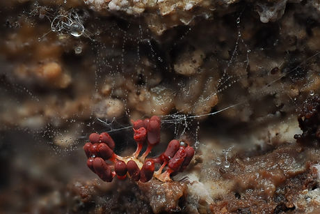
Leocarpus fragilis - has distinctive, short-stalked to near sessile, crowded, ellipsoid to subglobose sporangia (> 4mm high) which in the following photo looks more like the contents of a tin of red-kidney beans rather than the baked beans appearance of the previously featured Oligonema spp. However, the colour actually varies from yellow-brown to chestnut brown or deep maroon so a degree of caution is required, but in conjunction with the short-cylindrical to pear-shaped, smooth, shiny peridium, they're unlikely to be misidentified; occurs in leaf-litter, especially of conifers, Birch and Gorse, often clustering on vegetation or up the trunk of small trees [more photos required].

Leocarpus fragilis
Physarum - with around fifty described species, it's hard to know where to start with this genus. However, many appear to be either extremely rare or under-recorded so, after looking at local distribution records and species that other photographers have found and have had positively identified, I believe that there are certainly two that I should be able to find and possibly three or four others that I might find at some point.
Physarum album [formerly Physarum nutans] - the most frequently recorded member of the genus; stalked subglobose or lenticular, usually nodding, white or pale grey sporangia (> 1.5mm tall); peridium encrusted with lime, the upper portion splitting into irregular fragments or petaloid lobes, base persistent, spores chocolate-brown in mass; stalk long, tapering, longitudinally wrinkled, darker below, paler towards the apex; distinctive due to the drooped head covered in white crystals, and later with petaloid dehiscence; gregarious typically in large spaced groups; occurring on deadwood, and sometimes on bark of living trees [best photographed before dehiscence].
Physarum leucophaeum - stalked, erect, subglobose, white or bluish-grey sporangia (> 1.5mm tall); peridium, thin, usually thickly covered with calcareous granules, thickened base persistent as a shallow cup, spores black in mass; stalk often twisted, mid-brown to reddish-brown, powdered with white; occurs on rotten wood [best photographed before dehiscence].
Other species that I'd like to find include Physarum compressum, Physarum psittacinum and Physarum viride.
.jpg)
Physarum sp. (still developing)

Physarum album

Physarum album

Physarum album
-2.jpg)
Physarum album (spored)
.jpg)
Physarum album (spored)

Physarum cf.leucophaeum